Cerebrovascular Tests
Diagnostic Imaging Techniques for Vascular Malformations
Accurate diagnosis and detailed imaging information about a patient’s cerebrovascular disease, based on the advanced imaging techniques, selected according to each patient’s particular situation, are computed tomographic imaging (CT) and angiography (CTA), magnetic resonance imaging (MRI) and angiography (MRA), and angiogram arteriogram:
Computed tomographic imaging (CT) and angiography (CTA):
This noninvasive imaging technique requires lying very still on your back in a tubular machine that uses X-rays and a special dye injected through an IV in your arm to look closely at blood vessels in the brain.

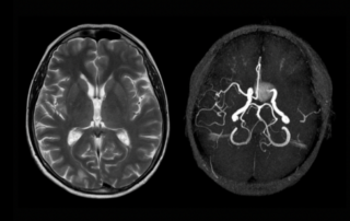
Magnetic Resonance Imaging (MRI); Magnetic Resonance Angiography (MRA):
Additional noninvasive tools, MRI and MRA imaging also require lying face up and motionless in a tube-shaped machine that uses magnetic fields and a special dye injected through an IV in the arm to look closely at the blood vessels of the brain.

Cerebral Angiogram
For this imaging technique, the patient is sedated and a small area of the body — typically the groin — is numbed. Then, one of the endovascular neurosurgeons or neuro-interventionists inserts a small tube, called a catheter, into a blood vessel and threads it up to the neck and brain, where dye is injected so that X-rays can reveal all arteries and any abnormalities. When the angiogram has been completed, the patient remains in the hospital, lying flat, for two to six hours to help prevent post-procedure complications. The length of time each patient remains lying down depends on the type of closure used and on the particular patient.