Arteriovenous Malformations
What are Arteriovenous Malformations (AVMs)
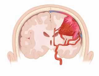
AVMs are abnormal tangles of blood vessels within the brain — called arteriovenous malformations because abnormal arteries are connected to abnormal veins. The abnormality is congenital (present from birth). It is found in 1 in 700 people (0.14 percent of the general population). Almost all AVMs are non-hereditary (do not run in families). Click for Images

Symptoms
Many people who have an AVM experience no symptoms at all and are diagnosed by chance when they visit a doctor about a different concern. When AVMs cause physical problems, patients are likely to experience one or more of four main symptoms:
Seizure
While AVMs can cause seizures, not all result in the type of the seizure in which the body shakes rapidly and uncontrollably. There are many types of seizures; some have mild symptoms, last from 30 seconds to two minutes and do not cause lasting harm. Others last longer or can occur one after the other.
Headache
Although many patients with an AVM have headaches, the vast majority of the pain proves to be unrelated to the AVM. Yet the AVM itself can cause headaches in a small number of patients, and these can fade when the AVM is treated.
Stroke-like effects
In some patients, blood flow is diverted away from part of the brain to the AVM, which can lead to symptoms similar to those caused by a stroke. A patient may suffer weakness or numbness on one side of the body, difficulty speaking or understanding speech, loss of vision, double vision and balance difficulties.
Brain hemorrhage
AVMs can rupture and cause bleeding in the brain (also called a bleeding, or hemorrhagic, type of stroke). Symptoms that suggest brain hemorrhage include sudden onset of a severe headache; nausea, vomiting, weakness or numbness on one side of the body; difficulty speaking or understanding speech; loss of vision; double vision and balance difficulties. If you experience any of these symptoms, call 9-1-1. Brain hemorrhage is a serious form of stroke that can cause serious brain injury and even death, although many patients make good long-term recoveries.
Treating AVMs
Decisions about treatment are made between the patient and a team of doctors on a case-by-case basis and depend on the location and size of the AVM, the severity and nature of the symptoms, the patient’s age and health status, and the risk involved in treatment. Treatment options for brain AVMs include:
Observation
This may be an appropriate approach for certain patients, including people who are older, have multiple medical problems or have complex AVMs for which treatment carries high risk.
Surgery
In this inpatient procedure, the AVM is removed from the brain through open surgery. If successful, it provides immediate protection against AVM rupture. Surgery is a good option for many AVM patients; however, if the AVM is complex or difficult to reach, this treatment may be a less attractive option.
Gamma Knife® Radiosurgery
This less-invasive form of treatment is a one-day outpatient procedure in which beams of radiation are precisely focused on the AVM, causing it to shrink over time. In most patients, the AVM will be cured in one to three years following treatment. Such radiosurgery is most useful for smaller AVMs, but can be used selectively in certain patients whose AVMs are larger.
Endovascular Embolization
A small catheter (tube) is used in this inpatient procedure. The catheter is passed from a blood vessel in the groin (or arm) up into the AVM, where glue or other material is injected. Endovascular embolization is primarily used in preparation for surgery or radiosurgery to make the treatments safer and more effective. In some cases, the treatment can cure the AVM, making surgery or radiosurgery unnecessary.
Online Resources for Patients
The Aneurysm and AVM Foundation